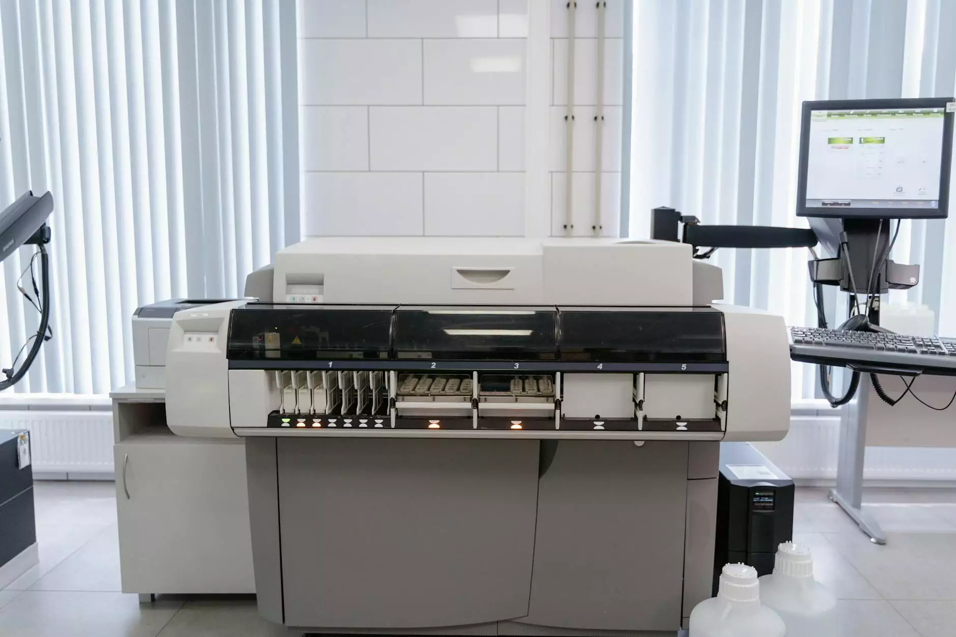
The Western Blot Imaging System: An Essential Tool for Molecular Biology

The field of molecular biology has witnessed tremendous advancements over the years, allowing scientists to dig deeper into the complexities of cellular functions and interactions. One of the pivotal techniques at the forefront of these innovations is the Western Blot Imaging System. This sophisticated tool not only assists in detecting specific proteins in a sample but also plays a crucial role in various research disciplines, including immunology, biochemistry, and cell biology. In this article, we will explore the components, functionalities, and the far-reaching implications of the Western Blot Imaging System in scientific research.
What is the Western Blot Imaging System?
The Western Blot Imaging System is a powerful analytical tool used to detect and quantify specific proteins present in a biological sample. The process involves several key steps, including:
- Sample Preparation: Proteins are extracted from cells or tissues and then separated based on their size through gel electrophoresis.
- Transfer: The separated proteins are transferred onto a membrane, typically made of nitrocellulose or PVDF, which provides a solid support for further analysis.
- Blocking: To avoid non-specific binding, the membrane is incubated with a blocking solution containing proteins that saturate potential binding sites.
- Antibody Incubation: First, a specific primary antibody is added that binds to the target protein, followed by the addition of a secondary antibody conjugated to a detectable marker.
- Imaging: The Western Blot Imaging System captures images of the bands representing the proteins, allowing researchers to analyze their presence and quantity.
This intricate process allows for the precise analysis of protein expression levels, post-translational modifications, and interactions between proteins, making it invaluable in molecular research.
Key Components of the Western Blot Imaging System
A state-of-the-art Western Blot Imaging System consists of several essential components, each contributing to the overall accuracy and efficiency of the protein analysis. These include:
- Gel Electrophoresis Apparatus: This equipment is crucial for the separation of proteins based on size. High-quality gel rigs ensure proper resolution of bands.
- Transfer Systems: Efficient transfer systems maintain the integrity of proteins during the transition from gel to membrane.
- Membranes: Nitrocellulose and PVDF membranes are commonly used due to their excellent binding properties and ability to withstand various detection methods.
- Antibodies: The choice of antibodies is vital; researchers must select high-affinity antibodies to ensure specific binding to the target protein.
- Imaging Software: Advanced imaging systems utilizing software for quantification and analysis enable accurate interpretation of protein levels.
- Detection Systems: This includes chemiluminescent or fluorescent markers that allow visual detection of the proteins on the membrane.
Applications of the Western Blot Imaging System
The versatility of the Western Blot Imaging System means it has found applications across various disciplines in biological research. Some notable uses include:
1. Disease Diagnosis
Several diseases, especially autoimmune disorders and viral infections, can be diagnosed through protein detection in patient samples. For instance, the presence of specific antibodies in the blood can indicate conditions like HIV and Lyme disease.
2. Cancer Research
In cancer studies, Western blotting assists in analyzing the expression of oncogenes and tumor suppressor proteins, providing crucial insights into the mechanisms of tumorigenesis.
3. Protein Interaction Studies
Understanding protein interactions is fundamental to deciphering cellular pathways. Western blotting can identify co-immunoprecipitated proteins, revealing potential interactions.
4. Drug Development
Western blot analysis is essential in evaluating the efficacy of new drugs by monitoring changes in protein expression levels after treatment. This helps in understanding the mechanism of action of pharmaceuticals.
Advantages of the Western Blot Imaging System
Utilizing the Western Blot Imaging System comes with numerous advantages that enhance the reliability of protein analysis:
- Specificity: The use of highly specific antibodies allows for accurate detection of target proteins amidst a complex background.
- Sensitivity: Modern imaging systems provide high sensitivity, enabling the detection of proteins present in low abundance.
- Quantification: The capability to quantify proteins allows researchers to assess relative expression levels, crucial in many studies.
- Visual Documentation: Permanent records of results via imaging facilitate better data management and sharing among researchers.
- Cost-Effectiveness: Compared to other protein detection methods, Western blotting is relatively inexpensive and does not require extensive equipment.
Best Practices for Using a Western Blot Imaging System
To maximize the effectiveness of the Western Blot Imaging System, researchers can adhere to best practices that enhance accuracy and reproducibility:
1. Sample Preparation
Proper sample preparation is crucial. Ensure consistency in sample collection, handling, and storage to minimize variability in results.
2. Antibody Validation
Always validate the specificity and sensitivity of the primary and secondary antibodies used. This step is crucial for ensuring reliable results.
3. Optimization of Transfer Conditions
Optimize transfer conditions such as time and voltage to ensure complete transfer of proteins while preventing degradation.
4. Standardization of Detection Methods
Standardize your detection method by using controls, which can help establish consistency across experiments.
The Future of Western Blot Imaging Systems
The future of the Western Blot Imaging System looks promising, with continuous advancements expected in technology that will enhance its capabilities:
- Automation: The trend toward automation will streamline processes, reduce human error, and increase throughput in laboratories.
- Integration with Other Techniques: Combining Western blotting with techniques like mass spectrometry can provide deeper insights into protein functions and interactions.
- Smart Imaging Systems: The development of intelligent imaging systems utilizing machine learning algorithms may improve analysis speed and accuracy.
As technology progresses, the Western Blot Imaging System will continue to be an indispensable tool in the arsenal of molecular biologists, facilitating groundbreaking research and discovery.
Conclusion
In summary, the Western Blot Imaging System is an essential technique in molecular biology that provides profound insights into the proteome. By allowing researchers to detect and quantify specific proteins, this system has significantly advanced our understanding of various biological processes and disease mechanisms. As we look to the future, embracing innovations in this field will undoubtedly enhance the efficacy and applications of the Western blotting technique, reinforcing its status as a cornerstone of protein analysis.


